Neurological consultation and evaluation with Neurologist
We start each case with consultation from a board-certified neurologist which includes detailed review of symptoms, signs, prior treatment trials, tests and imaging, results. Our neurologists also closely examine and discuss findings on brain mapping and digital cognitive exams, fMRI and any other diagnostics imaging in detail. Following careful review of all the above data, our team designs an individualized treatment protocol and discusses the recommendations with you in office.
Quantitative Electroencephalogram (QEEG) and ‘Brain Mapping’
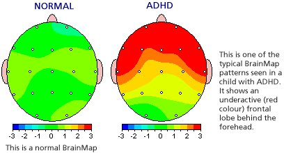
QEEG is an advance form of evaluating individual brain regions by collecting and analyzing the electrical ‘brain waves’ (e.g. frequency and amplitude) and digitally comparing the data against a database of age-matched otherwise health individuals. This brain map is a very precise evaluation that can help 1) make more precise diagnoses as each disorder often has a ‘signature’ pattern on brain map; and 2) help design treatment protocol to reverse the areas of abnormality to a normal state. In addition, brain mapping can help patients visualize the exact abnormalities and see the ‘before’ and ‘after’ results of treatment tab. Learn more from our Real Cases.
NeuroTrax Cognitive Exams
NeuroTrax is a battery of scientifically-validated cognitive tests that evaluate ‘brain fitness’ including executive function, working memory, visual spatial skills, verbal fluency, and attention span. This advanced computerized assessment helps establish baseline status and show progress over the course of treatment. Patients will be able to establish goals, be it improved attention, memory, or executive function and clearly visualize their achievements.
MRI and functional fMRI
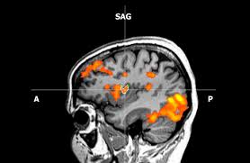
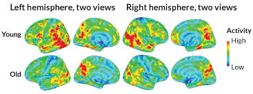
In addition to consultation, brain mapping, and digital cognitive exams, our team recommends either anatomical MRI or functional MRI imaging. The MRI imaging is done at specialized centers that have the required expertise and scanning technology to enable precise navigation to chosen brain targets when performing transcranial magnetic stimulation (TMS). Functional MRI is also done to precisely evaluate the areas of abnormal blood flow (BOLD) and the status of connectivity between different networks of the brain. As each individual brain operates differently and each disorder is associated with different connectivity and blood flow changes, fMRI allows for far more precise and, often, more likelihood of successful treatment.
Neuronavigation
At most other centers, Transcranial Magnetic Stimulation (TMS) is often done without precise localization of target brain regions. Scalp measurements are used which, while acceptable, cannot match the precision and accuracy the MRI-guide neuronavigation brings. Currently, no other center in North America offers this specific type of navigated TMS treatment.


